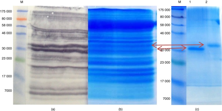
Figure 2.

Steps showing the identification and purification of two cross‐reactive peanut antigens. (a) Western immunoblot showing the identification of peanut antigens that are cross‐reactive with IgG antibodies in an anti‐Schistosoma mansoni soluble egg antigen (anti‐SmSEA) serum, (b) a portion of gel “a” stained in Coomassie blue for identification and excision of a pair of cross‐reactive peanut gel bands at ~30 000–33 000 MW intended for purification, (c) Coomasie‐stained SDS–PAGE showing further resolution of the pair of peanut bands into a lower band (lane 1) and a top band (lane 2) for mass spectrometry analysis. [Colour figure can be viewed at wileyonlinelibrary.com]
