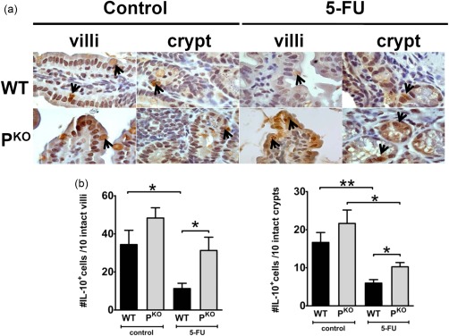
Figure 5.

Interleukin (IL)‐10 expression is detected in the epithelium. “Control” in the figure refers to mice which did not receive 5‐fluorouracil (5‐FU). (a) Immunohistochemical staining for IL‐10 shows that epithelial cell expression was reduced in inflamed wild‐type (WT) but not PKO mice. Isotype matched antibody or anti‐IL‐10 used on sections from IL‐10–/– mice did not demonstrate any staining (not shown). (b) Quantification of IL‐10 positive epithelial cells/10 crypts or villi (shown separately). Cells were counted from intact villi/crypts only. Data are shown as mean ± standard error of the mean (s.e.m.) (n = four or five mice/group). *P < 0·05 and **P < 0·01.
