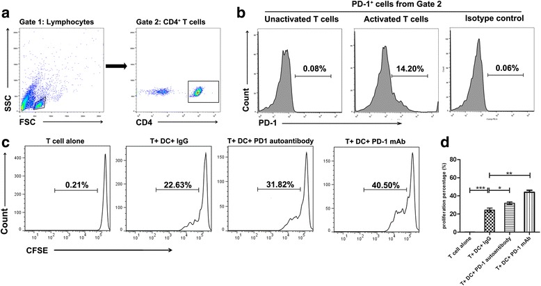
Fig. 4.

PD-1 autoantibodies increased T cell proliferation. Autologous T cells and DCs were co-cultured for 72 h. a, b PD-1 expression on T cells activated by anti-CD4 detected by flow cytometry. c The division of CFSE-labeled CD4+ T cells determined by flow cytometry. The data represent one of three independent experiments. d The statistic graph of three experiments. Values represent the means ± SD. *p < 0.05, **p < 0.01, ***p < 0.001. DCs dentritic cells, Ig immunoglobulin, mAb monoclonal antibody, PD-1 programmed cell death protein 1
