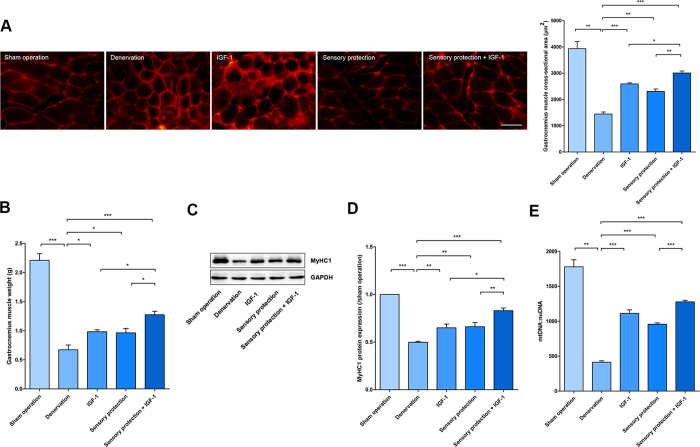
Figure 1. IGF-1 promoted the effects of sensory protection in vivo.
(A) Cryosections of GAS muscles were stained with dystrophin. Representative images are shown. Scale bar = 50 μm. Mean CSA of GAS muscles in the sham operation, denervation, IGF-1, sensory protection and sensory protection + IGF-1 groups at the 8th week after surgery (n = 5). (B) Mean GAS muscle wet weight for the different treatments. (C) The total protein extracted from GAS muscle samples was immunoblotted with antibodies against MyHC1 and GAPDH. GAPDH was used as a loading control. (D) Quantification of MyHC1 protein levels (n = 5). (E) Quantification of the number of mitochondria in GAS muscle cells (n = 5). Bar graphs with error bars showing the mean ± SEM. *P < 0.05, **P < 0.01, ***P < 0.001.

