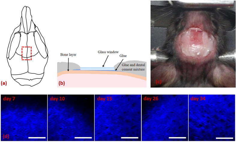
Figure 1. Mouse calvarial window model for longitudinal intravital BM microscopy.
(a) Schematic of mouse calvaria on which the position of the glass window is indicated (red dashed box). (b) Schematic showing how the glass window is attached to the mouse skull with glue. (c) Photograph of the mouse calvarial window. (d) SHG images of the bone surface at the same site on the calvaria taken 7, 10, 15, 26, and 34 days after the glass window was attached. Scale bar: 100 μm.

