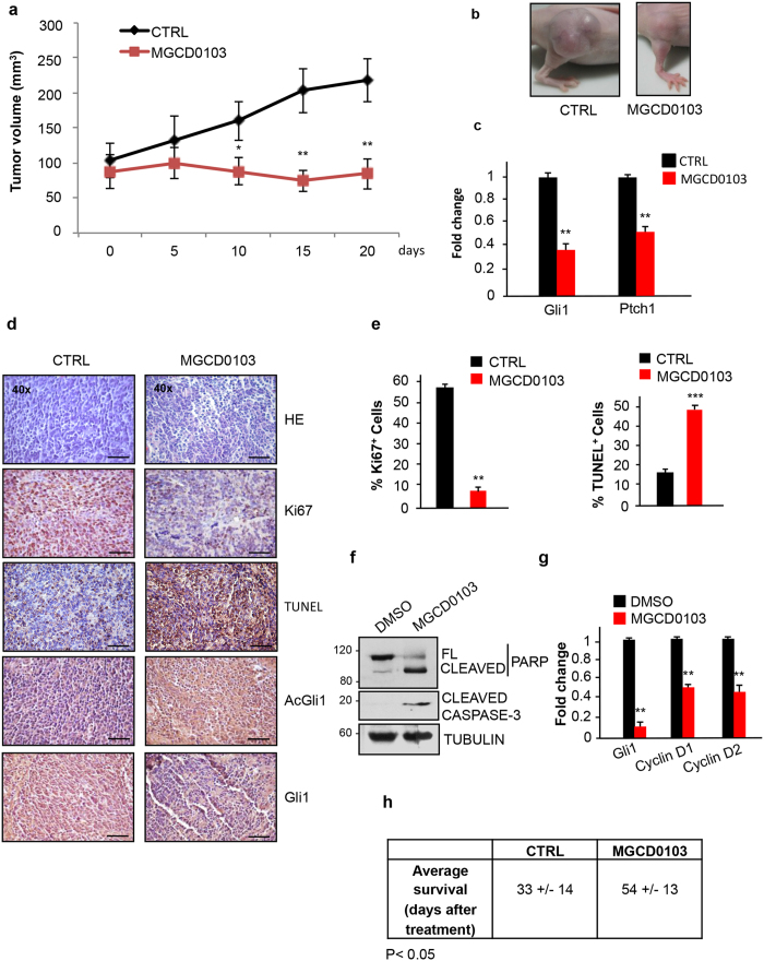
Figure 4. Pharmacological inhibition of HDAC1 and HDAC2 counteracts Shh-dependent medulloblastoma growth in vivo.
(a) Primary SHH-MB cells were grafted into nude mice (2 × 10⁶ cells/flank). After two weeks from the injection, when the tumor volumes reached 100 mm3, mice were treated daily with 170 mg/Kg MGCD0103 or H2O per os. The tumor masses were measured with a caliper at the indicated times. (n = 8 for each experimental group) (b) Representative pictures of tumor masses at the end of the experiment (CTR vs MGCD0103) (c) QPCR analysis of Gli1 and Ptch1 mRNA levels from pooled RNA samples extracted from the explanted tumor masses of each experimental group. Results were performed in triplicates. (d) Immunohistochemical staining of Gli1, AcGli1, Ki67, and Tunel assay in sections from the allografted tumors. Hematoxylin/Eosin staining was performed to control the quality of the slides (scale bar, 100 μm). (e) Analysis of the percentage of Ki67 positive cells and percentage of apoptotic cells from the sections in Fig. 4d. (f) Western blot analysis on MB samples (pools of 4 samples for each experimental group) excised from Math1-Cre/Ptcfl/fl mice subcutaneously treated for 6 hours with DMSO or MGCD0103 (170 mg/Kg). Apoptosis was evaluated by analyzing PARP and caspase 3 processing. Tubulin, loading control. (g) QPCR analysis of Gli1, Cyclin D1 and Cyclin D2 levels of MB samples from Fig. 4f (h) Average survival of Math1-Cre/Ptcfl/fl mice treated with MGCD0103 (80 mg/kg) or H2O per os starting from 4 weeks of age. MGCD0103 (n = 6) CTRL (n = 6). ***p < 0.001; **p < 0.01; *p < 0.05. Uncropped Western blot gels related to this figure are displayed in Suppl. Fig. 7.

