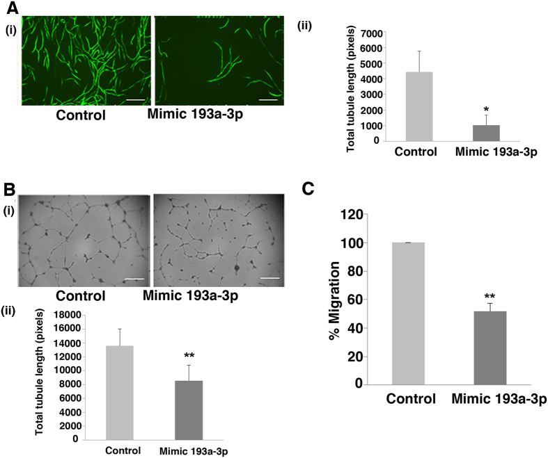
Figure 3. mir-193a-3p effect on CB ECFC-derived cell angiogenic functions.
(A) Transfected CB ECFC-derived cells were plated on hBM MSCs in a 96 well plate and images at x10 magnification were taken at Day 12 (endpoint). Effect of miR-193a-3p mimic on tubule formation of CB ECFC-derived cells were quantified using Angiosys software (n = 3, *p < 0.05). Scale bar, 100 μm. (B) Transfected CB ECFC-derived cells were plated onto growth factor reduced matrigel and allowed to migrate for 18 hr before images at x4 magnification were taken and quantified using Angiosys software (n = 3, p < 0.01). (C) CB ECFC-derived cells transfected with 10 nM miR-193a-3p mimic for 48 hr were suspended in 0.5% FBS in EBM-2 and placed in 8 μm transwells and allowed to migrate towards 10% FBS in EGM-2 growth media (**p < 0.01). The values presented are the mean ± S.E.M of three independent experiments (*p < 0.05; **p < 0.01; Student’s t-test).

