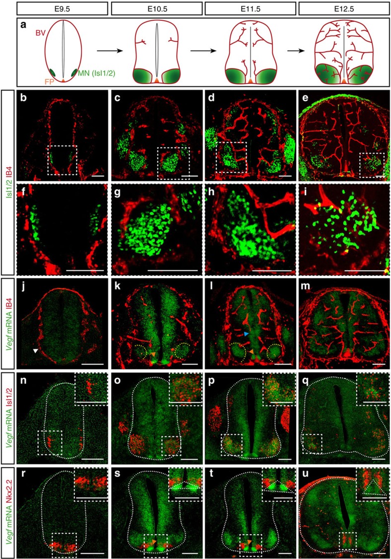
Figure 1. Blood vessel sprouts invade the developing mouse spinal cord at specific locations and grow in a stereotypical pattern.
(a) Scheme of SC vascularization during mouse development (E9.5 till E12.5), showing blood vessels (BV, red), the floor plate (FP, orange) and the MN columns (green). (b–e) Representative images of SCs at the developmental stages indicated, showing labelled endothelial cells (IB4+) and post-mitotic MNs (Isl1/2+). (f–i) Higher magnifications of insets in b–e. Note blood vessels stay outside the Isl1/2+ domain till E12.5. (j–m) Representative images of blood vessel staining (IB4+) in the SC combined with ISH for Vegf from E9.5 till E12.5 in mice. At E9.5 (j), Vegf is uniformly expressed in the entire SC (white arrowhead: PNVP). From E10.5 till E12.5 (k–m) Vegf expression becomes restricted to specific neuronal domains (yellow dotted lines: MN columns; orange arrowheads: FP; blue arrowhead: neuronal progenitors). (n–q) Representative images of ISH for Vegf combined with immunostaining for MNs (Isl1/2+) confirming that Vegf expression is highly localized and increased in MN columns from E10.5 onwards. Insets show higher magnifications of MN columns. (r–u) Representative images of ISH for Vegf combined with immunostaining for the p3 neuronal progenitor domain (Nkx2.2+) revealing expression of Vegf in the p3 domain and in the FP (orange arrowheads: FP (domain Nkx2.2−, below Nkx2.2+)). Insets show higher magnifications of the FP region (white dotted outlines: FP). Scale bars 100 μm.

