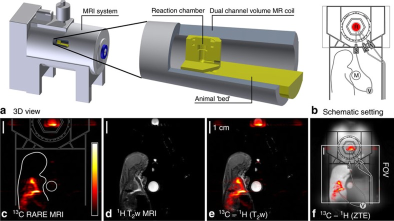
Figure 7. Simultaneous 13C-HP and ex vivo imaging:
The compact design of the setup enabled SAMBADENA next to a small rat post mortem in the sensitive volume of the magnet (a,b). Within seconds, the 13C-tracer HEP was polarized to ∼17% at a concertation of 22 mM. Without leaving the magnet, ∼330 μl of the tracer were injected into the rat, and strong 13C signal with a maximum SNR of 113 was observed by MRI acquired in 0.5 s (c). Subsequently, a T2-weighted 1H-MRI (d) and 1H Zero-Echo-Time (ZTE) MRI were recorded. Co-registration of 1H and 13C-data revealed the location of the tracer, in the reaction chamber and surrounding the heart of the animal (e,f). The isocenter of the magnet is the center of the images. R, reactor; FOV, field of view; M, Model Solution M3; 1, 2, 3: lines for pressure relieve, pH2 supply and pH2 injection, respectively.

