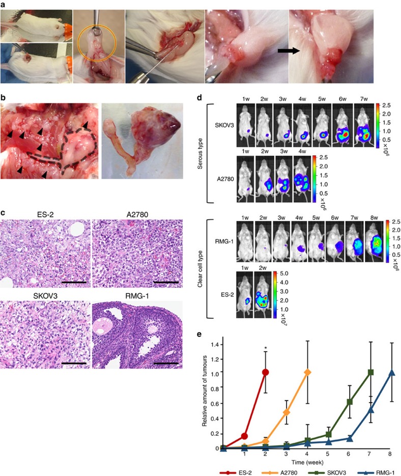
Figure 1. Establishment of mouse models for peritoneal dissemination in ovarian cancer.
(a) Illustrative photos of an orthotopic mouse model of ovarian cancer. A small incision (around 1 cm) was made on the left back of the mice. The ovaries were pulled out (red–yellow circle) and 1 × 10⁶ cells resuspended in 50 μl of PBS were injected into left ovarian bursa. (b) Representative photographs at the time of killing. The left photo shows metastatic tumours (black arrowheads) and the uterus with the primary left ovarian tumour (a black dotted line indicated the position), covered with fat pad. The right photo represents the dissected uterus with the primary left ovarian tumour. (c) Histological features of primary tumours in mouse model. The tumours were obtained from the orthotopic mouse model by using ES-2, A2780, SKOV3 and RMG-1 cells. Haematoxylin and eosin (H&E) staining. Scale bars, 100 μm. (d) Representative bioluminescence images of orthotopic mouse models. The mouse models were established by four types of ovarian cancer cell lines. Tumour progression was monitored weekly by an IVIS. (e) Quantitative analysis of tumour progression. To describe the differences in progression, the photon values were compared with the value at the time of killing, which was set to 1.0. n=3. Error bars represent s.d. There were significant differences at the 2 weeks time point by using analysis of variance followed by the Tukey–Kramer test for ES-2 versus other (*P<0.05).

