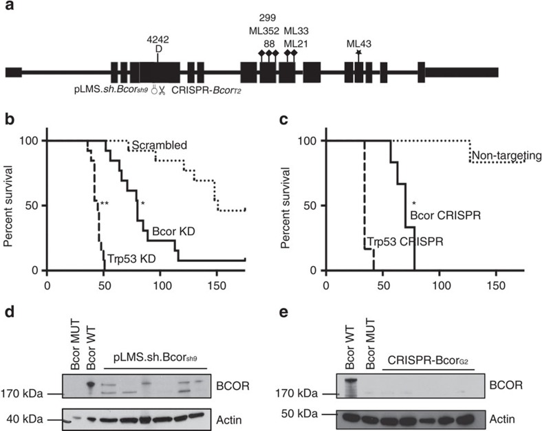
Figure 4. Experimental validation of BCOR tumour-suppressor function using RNAi and CRISPR-Cas9.
(a) Schematic showing exon structure of Bcor with position of identified spontaneous mutations and deletions and regions targeted using shRNA (pLMS-Bcor.sh9) or CRISPR-Cas9 (pCIG-BcorG2) guide RNA in exome 4. (b) Kaplan–Meier curve showing mice injected with 106 GFP+ve Eμ-Myc fetal liver cells (FLC) endowed with either pLMS.sh.Bcorsh9 (solid line), pLMS.shTrp531224 (heavy dashed line) or pLMS.sh.Scram (light dashed line). Mice transplanted with Eμ-Myc FLC endowed with pLMS.sh.Bcorsh9 (80 day median survival time post-transplant) or pLMS.shTrp531224 (45 day median survival time post-transplant) showed significantly accelerated lymphomagenesis compared with the cohort that received FLC transduced with pLMS.sh.Scram (151 day median survival time post-transplant). *=P value<0.05log-rank (mantel-cox) test, n=13 for each cohort. (c) Kaplan–Meier curve showing mice transplanted with 106 Eμ-Myc FLC endowed with either CRISPR-BcorG2 (solid line), CRISPR-Trp53 (heavy dashed line) or CRISPR-Scram (lightly dashed line). Mice transplanted with Eμ-Myc FLC endowed with CRISPR-BcorG2 (70 day median survival time post-transplant) or CRISPR-Trp53 (34 day median survival time post-transplant) showed significantly accelerated lymphomagenesis compared with the cohort that received FLC transduced with CRISPR-Scram. *=P value<0.05log-rank (mantel-cox test), n=6 for each cohort. (d) Immunoblot showing levels of BCOR knockdown in six tumours obtained from mouse recipients of Eμ-Myc pLMS.shBcor.sh9, #4242 (Bcor mutant) and #6066 (Bcor wild type). Whole cell lysates were prepared from Eμ-Myc-Bcor.sh9 fetal liver-derived tumours, a Bcor mutant Eμ-Myc lymphoma cell line (#4242) and a Bcor WT Eμ-Myc lymphoma cell line (6066). Western blot analysis was performed with antibodies specific to BCOR. Reduced BCOR protein expression was demonstrated in the Eμ-Myc pLMS.shBcor.sh9 lysates compared with WT. Equivalent protein loading was confirmed by probing for β-Actin. (e) Whole cell lysates were prepared from Eμ-Myc-pCIG-BcorG2 fetal liver-derived tumours, a Bcor mutant Eμ-Myc lymphoma cell line (4242) and a Bcor WT Eμ-Myc lymphoma cell line (6066). Western blot analysis was performed as in d above. No BCOR protein expression was apparent in the Eμ-Myc-pCIG-BcorG2 lysates compared with WT.

