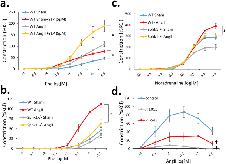
Figure 4. Contractile responses of the aorta and mesenteric arteries of WT and Sphk1−/− mice and effects of pharmacological inhibition of S1P pathway on vessel contraction.
(a) Tissue organ bath was performed using 2 mm aortic rings from WT mice preincubated for 5 min. with 5 μM S1P or with control buffer (n = 4/group); (b,c) Tissue organ bath was performed using 2 mm aortic rings (b) or 2nd order mesenteric arteries (c) from WT and Sphk1−/− mice with assessment of contractile force in response to Phe normalized to an initial KCl-induced contraction, n = 6/group; (d) Pharmacological studies on the effects of pre-incubation with 5 μM JTE-013 or 100 nM PF-543 on the contraction of WT Sham mesenteric arteries were performed using tissue organ bath, n = 4/group; *p < 0.05; †p < 0.05 as compared to control group. Phe = phenylephrine; Ang II = angiotensin II; [M] = molar concentration.

