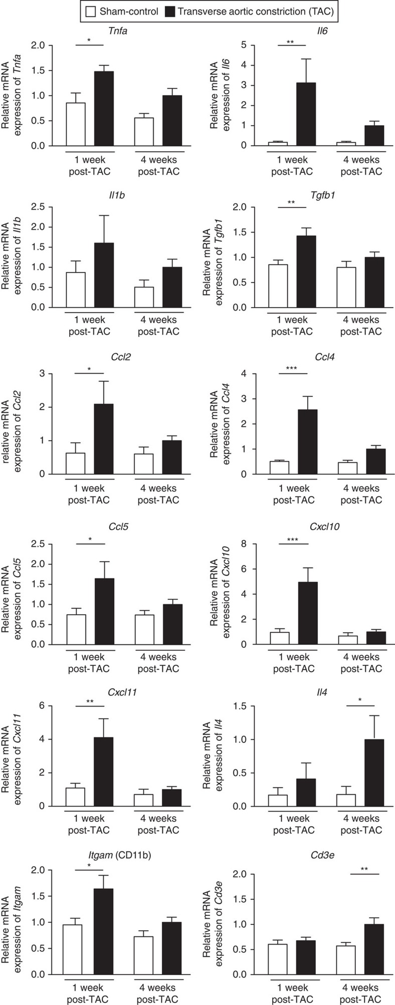
Figure 1. The inflammatory signature in hypertrophic left ventricle of mice.
Gene expression analysis (TaqMan real-time qPCR) of mediators of inflammation within the left ventricle of C57BL6/J mice. Relative mRNA expression in sham-operated control mice (white bars) and TAC-operated mice (black bars) at 1 and 4 weeks after surgery, internally normalized to 18 s ribosomal RNA expression. Tnfa, Il6, Tgfb1, Ccl2, Ccl4, Ccl5, Cxcl10, Cxcl11 and the innate cell marker Itgam (CD11b) were significantly increased in the TAC group compared with sham, 1 week after TAC. Four weeks after the operation, Il4 and the T cell marker Cd3e were significantly increased. Values are mean±s.e.m. (n=7–9). Two-way analysis of variance (ANOVA), Bonferroni post-test: *P value<0.05; **P value<0.01; ***P value<0.001.

