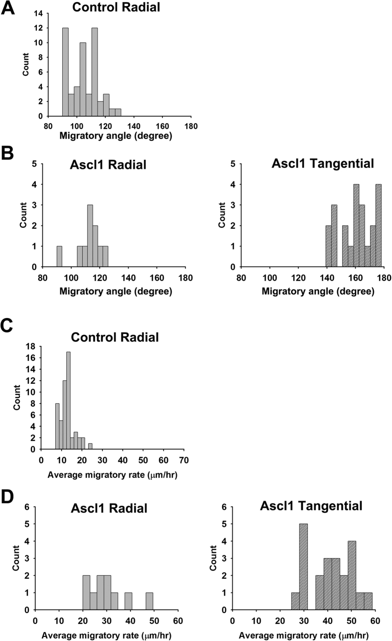
Figure 3. Overexpression of Ascl1 changes the migratory behavior of neurons in the dorsal telencephalon.
Ascl1 or US2 control expression construct were co-electroporated with a GFP expression plasmid. Two days after electroporation to the dorsal telencephalon, brains of E17.5 rats were dissected and sectioned in the coronal plane for slice culture and live imaging recording. The video from 5 to 11 hours after recording was used for tracking the migratory behavior. We set 0° to 180° axis in parallel to the lateral ventricle and the dorsomedial side as 180°. The line connecting the cell body location in the first frame (starting point) and the last frame (end point) of the video was used for measuring migratory angle. The average migratory rate was calculated as accumulated distance of every six-minute interval divided by recording time. 50 GFP-positive cells were counted for the control and 32 were counted for Ascl1 group. All counts were plotted into histograms according to their migratory angle or rate. (A) In the control group, most GFP-positive cells migrated radially in a migratory angle between 90° to 130°. (B) In Ascl1 group, GFP-positive cells were categorized into radially (90°–130°) and tangentially (140°–180°) migrating cells. (C) In the control group, GFP-positive cells migrated in the rate of 12.9 ± 3.6 μm/hour (mean ± SEM). (D) In Ascl1 group, radially migrating cells migrated in the rate of 30.2 ± 8.7 μm/hour and tangentially migrating cells migrated in the rate of 40.8 ± 9.3 μm/hour.

