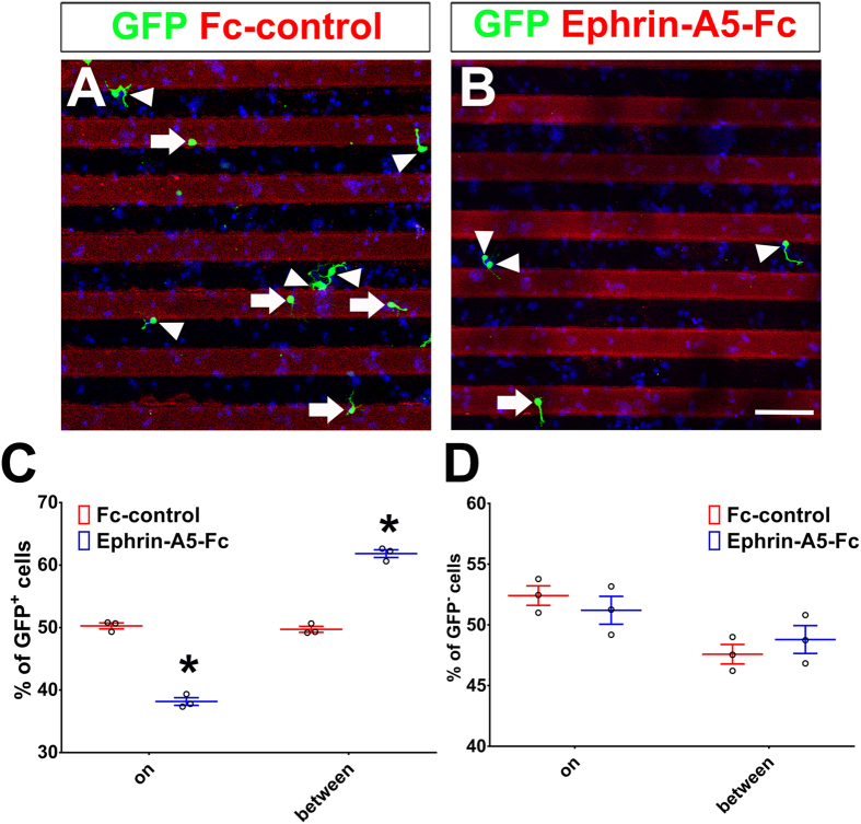
Figure 7. Ephrin-A5 has a repulsive effect on Ascl1-expressing cortical neurons.
Control (US2) or Ascl1 expression constructs were electroporated into the dorsal telencephalon of E15.5 rats. The dorsal telencephalon was dissected two days after electroporation and dissociated into individual cells. These cells were cultured on coverslips coated with Fc-control or Ephrin-A5-Fc stripes. Cells were fixed 16–18 hours after plating and cells on strips or between strips were counted. Electroporated cells were labeled with anti-GFP in green, nuclear DNA was stained with DAPI in blue. GFP-positive cells on stripes are indicated by white arrowheads; GFP-positive cells between stripes are indicated by white arrows. (A) On a coverslip coated with Fc-control, GFP-positive Ascl1-expressing cells and GFP-negative cells were distributed evenly on stripes and between stripes. (B) On a coverslip coated with Ephrin-A5-Fc, GFP-positive Ascl1-expressing cells were preferentially distributed between stripes, while GFP-negative cells were evenly distributed on stripes and between stripes. Length of the scale bar is 50 μm. (C,D) Distribution of GFP-positive and GFP-negative cells. 150 cells from each group were counted in each experiment. Data are presented as mean ± SEM with all data points and analyzed by using Chi-square test, n = 3. *p < 0.05 compared to the Fc-control.

