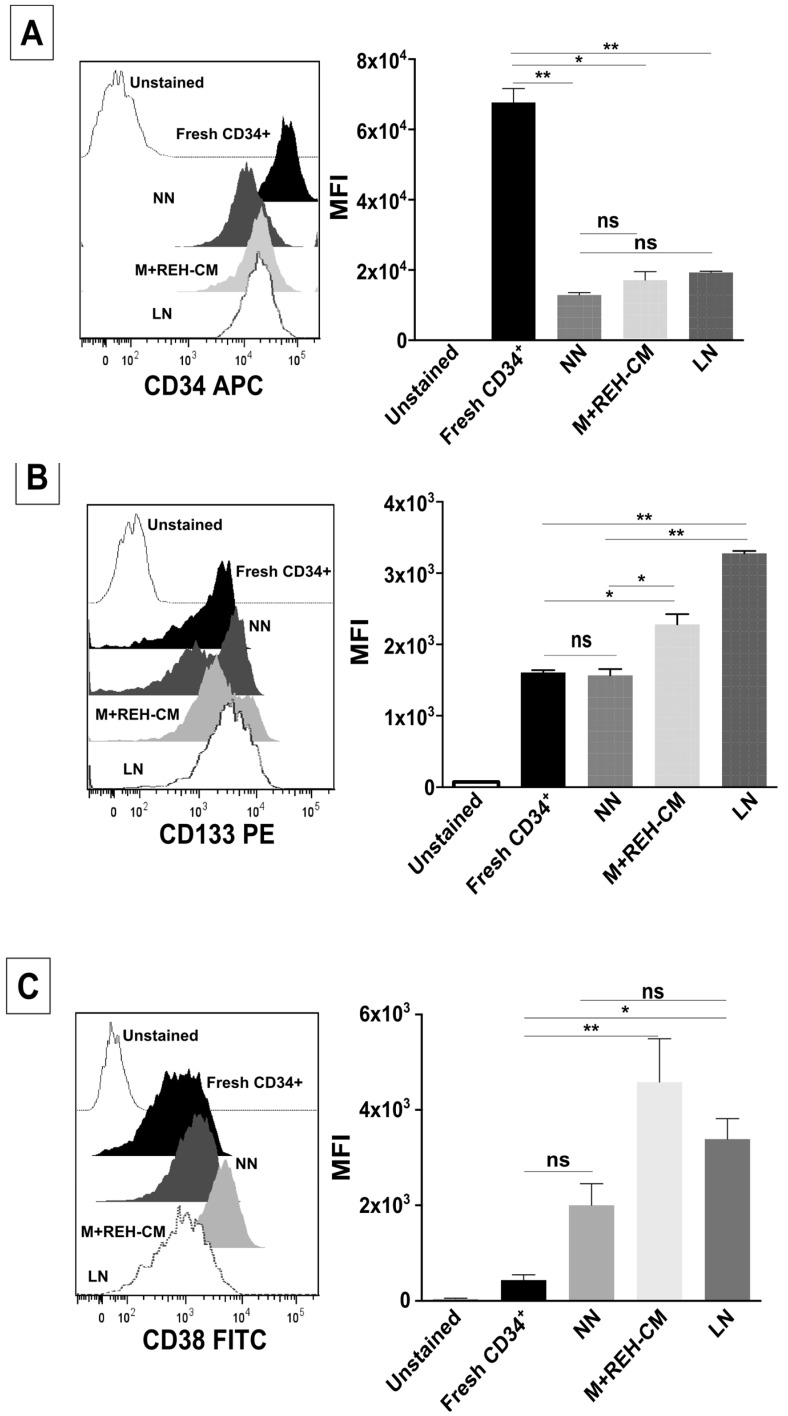
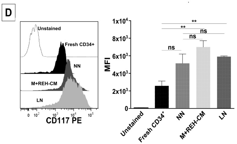
Figure 4.
Changes in stem cell and differentiation markers of CD34+ cells in the LN. Flow cytometry analysis of (A) CD34, (B) CD133, (C) CD38, and (D) CD117 (c-Kit) expression in freshly-isolated, NN, M+REH-CM, and LN CD34+ cells. Results are expressed as the median fluorescence intensity (MFI) obtained from two independent experiments done in triplicates (n = 6) (ns: non-significant, * p < 0.05, ** p < 0.01).


