Figure 6.
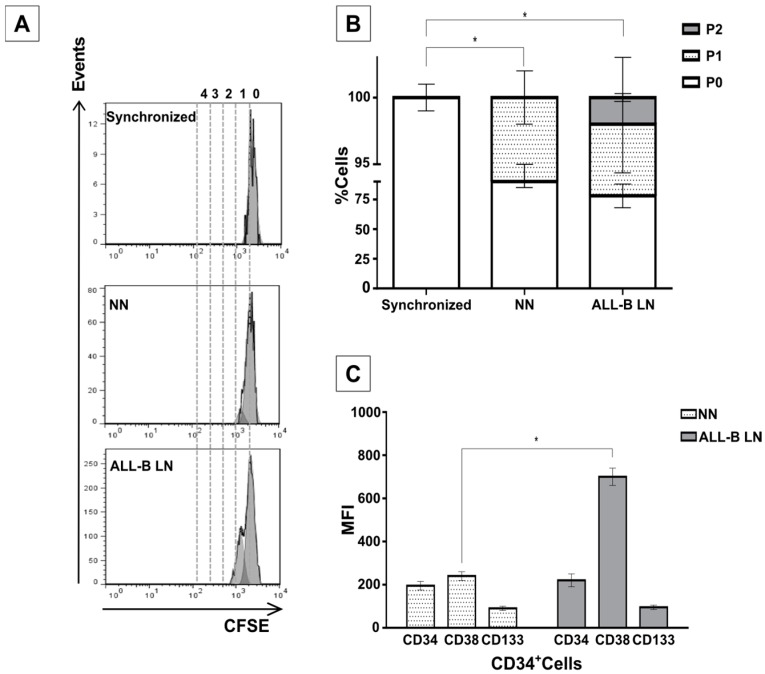
Increased proliferation and abnormal differentiation of CD34+ in a LN established with leukemic blasts from an ALL-B patient. (A) CD34+ cells isolated from an UCB sample and co-cultured with MSC pre-exposed to primary leukemic cells showed higher proliferation in the LN than in the NN. Synchronized cells are shown for comparison. A representative experiment is shown; (B) quantification of cells division in all settings are shown; and (C) mean fluorescence intensity (MFI) of the CD34, CD133 and CD38 markers in HSC co-cultured in the NN or in the ALL-B-LN. Data were obtained from two independent experiments done in duplicate (n = 4) (* p < 0.05).

