Figure 1.
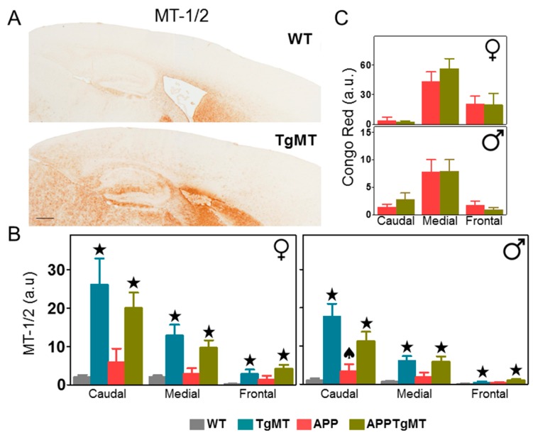
Effect of Mt1 overexpression on MT-1/2 and Congo Red staining in the cortex. (A) Representative brain MT-1/2 immunostaining in wild-type (WT) (top) and TgMT (bottom) mice; (B) Quantification of MT-1/2 IHC of the different genotypes in the cortex showed a dramatic increase in Mt1-expressing (TgMT and APPTgMT) mice (★ p at least ≤0.05 vs. WT or APP mice, respectively) with a prominent caudal-frontal gradient. As revealed by the significant interaction between APP expression and Mt1 overexpression (♠ p < 0.05 in male caudal region; the rest was not significant), APP expression tended towards an increase in MT-1/2 in WT mice; and the opposite was true in TgMT mice; (C) The greatest accumulation of dense amyloid plaques (stained with Congo Red) was localized in the medial area in both sexes. Results are mean ± SEM (n = 7–11). Scale bar: 400 µm. a.u., arbitrary units.

