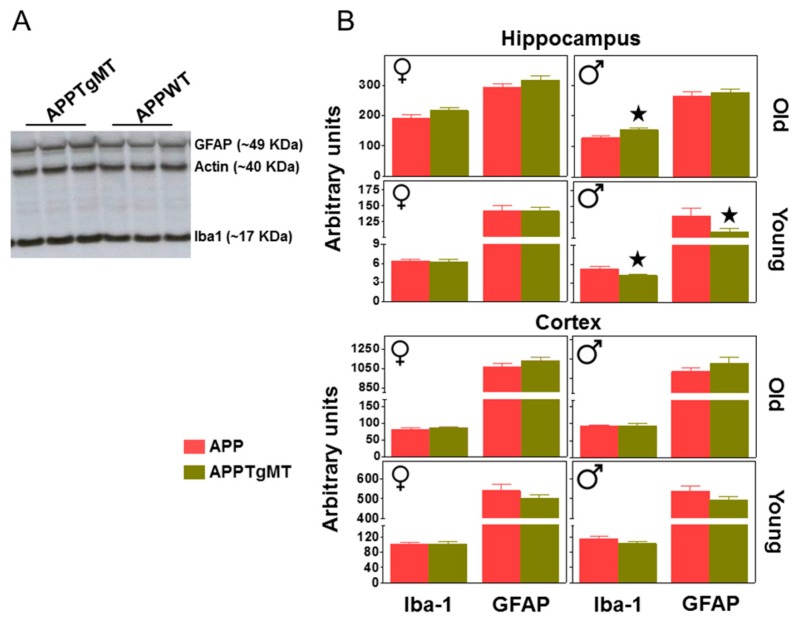
Figure 4.
Effect of Mt1 overexpression on hippocampal gliosis as measured by western blot (WB). Total hippocampal and cortex homogenates were assayed by WB to further characterize gliosis. (A) Representative band pattern of the WB (in an autoradiographic film) of old male hippocampus using antibodies for GFAP, Iba-1, and Actin; (B) Quantification of hippocampal GFAP and Iba-1 levels in young and old APPWT and APPTgMT mice. Iba-1 levels were increased by Mt1 overexpression in old male mice but decreased in young males; the latter also showed decreased GFAP levels. Data are mean ± SEM (n = 10–11). ★ p at least ≤0.05 vs. APPWT mice. a.u., arbitrary units.

