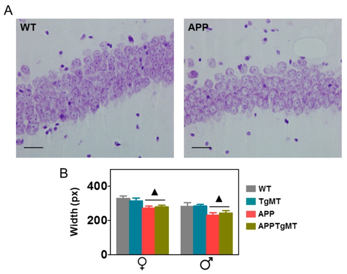
Figure 5.
Effect of Mt1 overexpression on hippocampal CA1 neurons. (A) Representative histochemistry of Nissl body staining of neurons in hippocampal CA1 of WT and APPWT mice. Scale bar: 20 μm; (B) Quantification of the thickness of the CA1 layer indicated a significant decrease in APPWT and APPTgMT mice in both sexes, whereas no significant effects of Mt1 overexpression were observed. Results are mean ± SEM (n = 11–18); ▲ p < 0.01 vs. APP negative mice.

