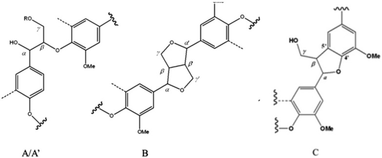
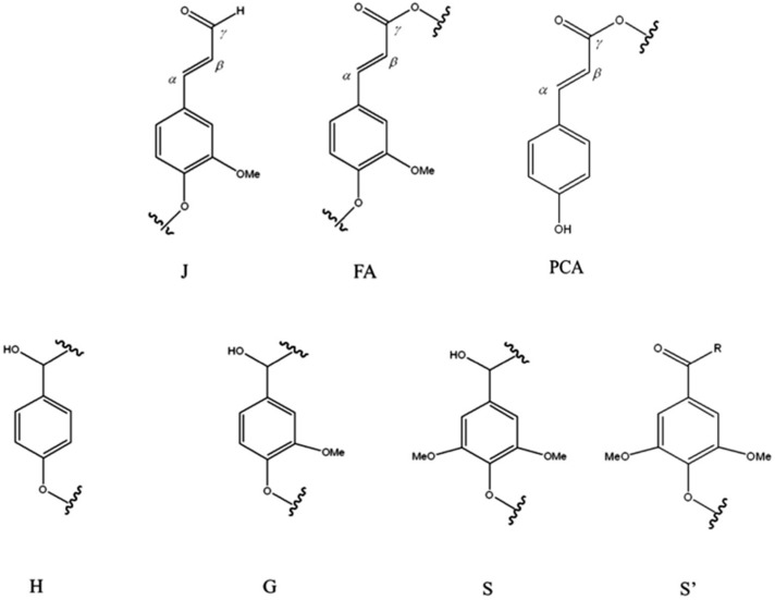
Figure 7.
Main molecular structures identified in wheat and Chamaecytisus proliferus lignin extracts by 13C-1H 2D-HSQC NMR spectroscopy. (A) β-O-4′ linkages; (A′) β-O-4′ linkages with acetylated γ-carbon; (B) resinol β-β′ structure; (C) phenylcoumaran β-5′ subunit; (H) p-hydroxyphenyl unit; (G) guaiacyl unit; (S) syringyl unit; (S′) oxidized syringyl units with a Cα ketone; (FA) ferulate; (PCA) p-coumarate and (J) cinnamyl aldehyde end-groups.


