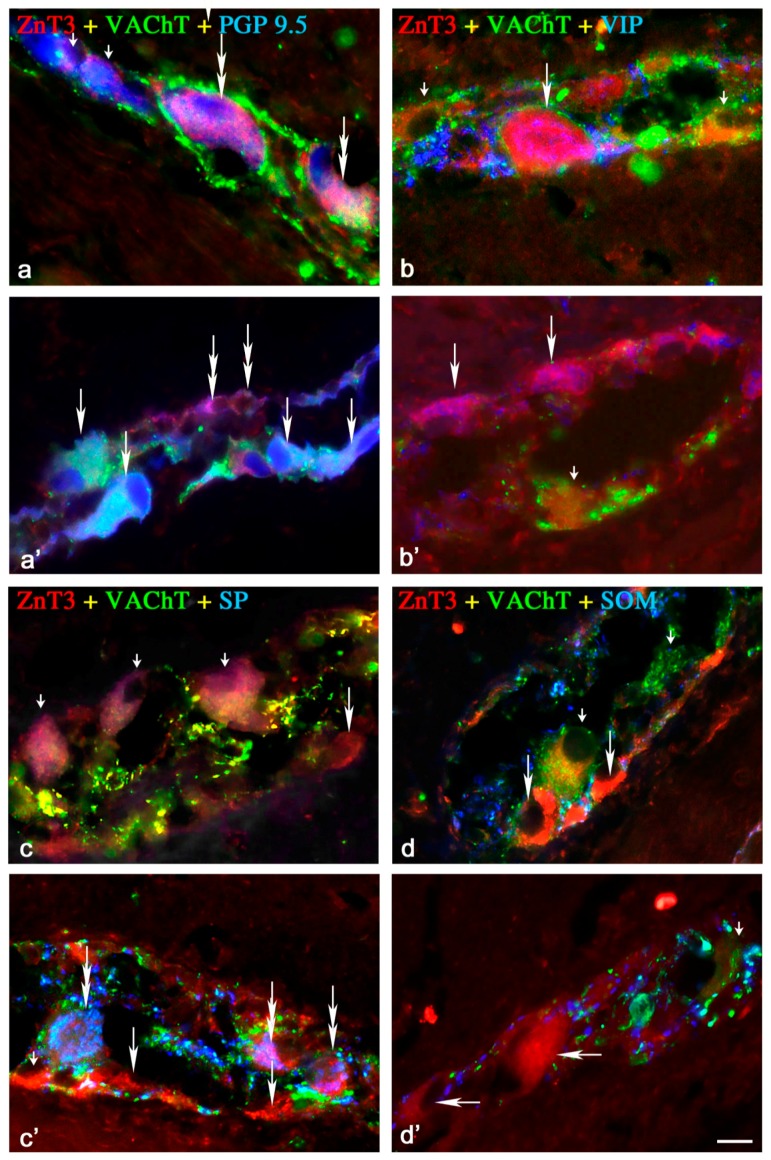
Figure 1.
Representative images of ZnT3+ neurons located in the myenteric plexus (MP) of the porcine ileum: myenteric ganglia. All images are composites of merged images taken separately from blue, red and green fluorescent channels. Control (C) group: (a) ZnT3+/VAChT+/PGP9.5+ neurons are indicated with double-headed arrows, ZnT3−/VAChT−/PGP9.5+ neurons are indicated with small arrows; (b) ZnT3+/VAChT+/VIP+ neurons are indicated with an arrow, ZnT3+/VAChT+/VIP− neurons are indicated with small arrows; (c) ZnT3+/VAChT+/SP− neurons are indicated with small arrows; ZnT3+/VAChT−/SP− neurons are is indicated with arrows; (d) ZnT3+/VAChT+/SOM− neuron is indicated with a small arrow; ZnT3+/VAChT−/SOM− neurons are indicated with arrows. Inflammatory (I) group (a’) ZnT3+/VAChT+/PGP9.5+ neurons are indicated with arrows, ZnT3+/VAChT−/PGP9.5+ neurons are indicated with double-headed arrows; (b’) ZnT3+/VAChT−/VIP+ neurons are indicated with arrows; ZnT3+/VAChT−/VIP− neurons are indicated with a small arrow; (c’) ZnT3+/VAChT+/SP− neuron is indicated with a small arrow; ZnT3+/VAChT−/SP+ neurons are indicated with double-headed arrows; ZnT3+/VAChT−/SP− neurons are indicated with arrows; Control group (d’) ZnT3+/VAChT+/SOM− neuron is indicated with a small arrow; ZnT3+/VAChT−/SOM− neurons are indicated with arrows. Scale bar 25 µm.

