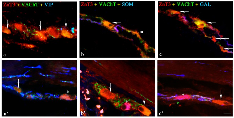
Figure 2.
Representative images of ZnT3+ neurons located in the outer submucous plexus (OSP) of the porcine ileum. All images are composites of merged images taken separately from blue, red and green fluorescent channels. Control (C) group: (a) ZnT3+/VAChT+/VIP− neurons are indicated with arrows; (b) ZnT3+/VAChT+/SOM− neurons are indicated with arrows; (c) ZnT3+/VAChT+/GAL− neurons are indicated with arrows; ZnT3+/VAChT−/GAL− neuron is indicated with a double-headed arrow. Inflammatory (I) group: (a’) ZnT3+/VAChT+/VIP− neuron is indicated with an arrow; ZnT3+/VAChT−/VIP+ neuron is indicated with a small arrow; (b’) ZnT3+/VAChT−/SOM+ neuron is indicated with a small arrow; ZnT3+/VAChT+/VIP− neurons are indicated with arrows; (c’) ZnT3+/VAChT−/GAL+ neuron is indicated with a small arrow; ZnT3+/VAChT+/GAL− neuron is indicated with an arrow. Scale bar 25 µm.

