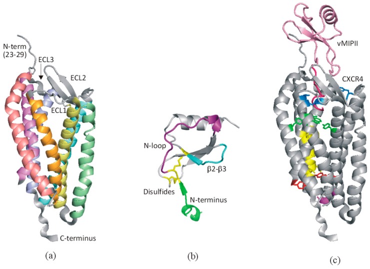
Figure 3.
Structural basis of chemokine-receptor recognition. (a) One monomer unit of the receptor CXCR4 (PDB code 4RWS [14]) with extracellular regions labeled; transmembrane helices are colored salmon (I), orange (II), yellow (III), green (IV), turquoise (V), violet (VI), and magenta (VII). (b) A typical chemokine monomeric unit (CCL2/MCP-1, PDB code 1DOK [18]) highlighting the critical regions for receptor recognition. (c) Structure of CXCR4 bound to vMIPII (PDB code 4RWS [14]) showing the chemokine in pink (N-terminal region in hot pink) and the receptor in gray, with residues proposed to be involved in transmembrane signaling [19] colored according to their putative roles: blue, chemokine engagement; green, signal initiation; yellow, signal propagation; red, microswitch residues; magenta, G protein coupling. In panels (a,c) residues 1-22 are not shown as they were not modeled in the crystal structure.

