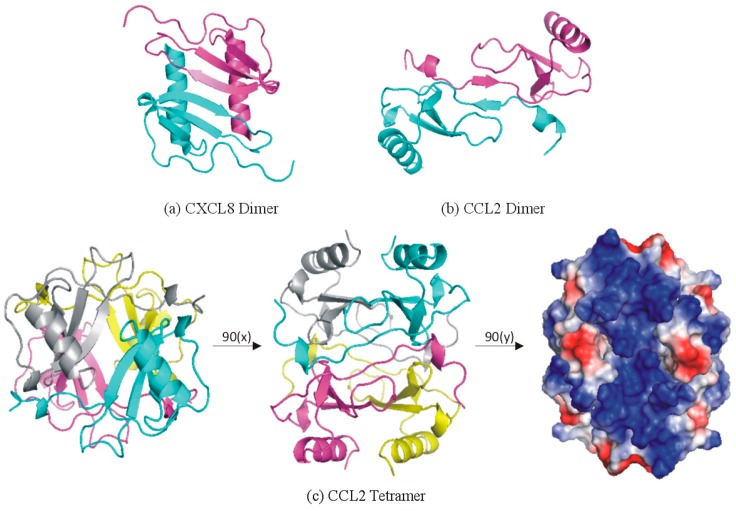
Figure 5.
Oligomeric structures of chemokines. (a,b) Dimer structures of (a) CXCL8/IL-8 and (b) CCL2/MCP-1, highlighting the distinct dimer interfaces for CXC and CC chemokines, respectively. (c) Tetramer structure of CCL2, highlighting: (left) the CXC-type dimer interfaces (cyan to gray and magenta to yellow protomers); (center) the CC-type dimer interfaces (cyan to magenta and yellow to gray protomers); and (right) the highly electropositive (dark blue) surface involved in GAG binding.

