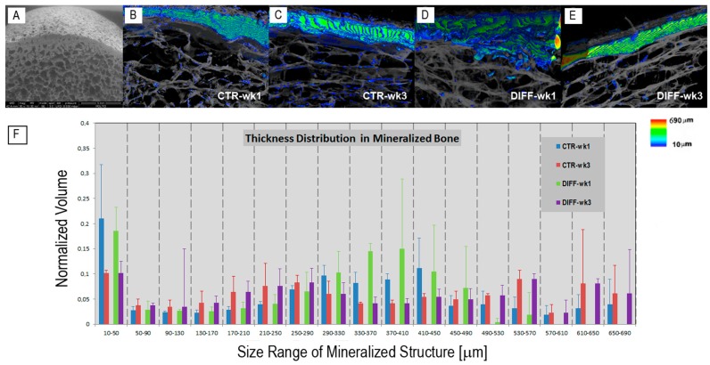
Figure 1.
(A) Scanning electron microscopy image of the Osteobiol® Dual Block before cell seeding: the cortical bone is anchored to cancellous bone, mimicking the natural human bone architecture; (B–E) Dual Block cultured with human Periodontal Ligament Stem Cells in xeno-free media. Synchrotron Radiation Phase-Contrast Microtomography (SR-PhC-microCT) three-dimensional (3D) images of sampling sub-volumes in (B,C) basal; and (D,E) osteogenic conditions. The cross-talk between cells, media, and scaffold produced 3D microCT images with two different phases, corresponding to different δ (refractive index decrement) values: the phase corresponding to demineralized Dual Block (DB) scaffolds (rendered in translucent gray) and the phase due to the contrast produced by the newly formed mineralized bone (colored in agreement to the color map of bone thickness distribution on the right); (B,D) at week 1; (C,E) at week 3; (F) Histograms of the fully mineralized bone thickness distribution in the trabecular portion of the investigated samples at weeks 1 and 3 of culture.

