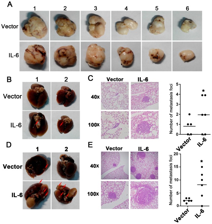
Figure 3.
Overexpression of IL-6 increases tumor metastasis in vivo. (A) Representative image showing subcutaneous tumors from the vector (upper panel) and IL-6 overexpression (lower panel) groups; (B) Representative image of tumor foci on the mice lung surface; Red arrows: tumor foci on the mice lung surface; (C) H & E staining of mice lung slices shows that IL-6 overexpression increased the metastatic capacity of breast cancer cells in vivo. The number of metastasis foci in the IL-6 overexpression group is more than that in the vector group, * represents p < 0.05; (D) Representative image shows the tumor foci on the lung surface of mice by tail vein injection; Red arrows: tumor foci on the mice lung surface; (E) Using the tail vein injection method, H & E staining of the lung slices shows that IL-6 overexpression in tumors increased the metastatic capacity in vivo. The number of metastasis foci observed under a microscope shows that the IL-6 overexpression group has more foci than the vector group, p < 0.05.

