Figure 1.
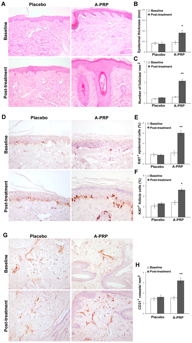
A-PRP treatment increased proliferation of epidermis basal cells and follicular bulge cells as well as vascularization. (A) Representative microphotographs of hematoxylin and eosin stained tissue sections from placebo and A-PRP treated scalp biopsies at baseline and two weeks post-treatment (original magnification 100×); (B,C) Morphometric evaluation of epidermis thickness (mm) and hair follicle density (no follicles mm−2); (D) Representative microphotographs of Ki67+ immunostaining of scalp biopsies from placebo and A-PRP treated patients at baseline and two weeks post-treatment (original magnification 200×); (E,F) The percentage of proliferating Ki67+ epidermal and follicle cells (dark brown nuclei); (G) Representative microphotographs of CD31 immunostaining of scalp biopsies from placebo and A-PRP treated patients at baseline and two weeks post-treatment (original magnification 100×); (H) Capillary density (CD31+ vessels mm−2). * and ** indicates p < 0.05 and p < 0.01, respectively.

