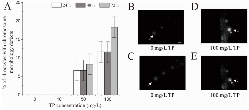
Figure 9.
Effects of exposure to TP for 4 h on the chromosome of −1 oocytes in diakinesis: (A) percent of −1 oocytes with chromosome morphology defects; (B,C) the chromosome of the same −1 oocyte in diakinesis of the control group on different focal planes, with six intact DAPI-stained bodies distributed dispersedly in the nucleus; and (D,E) the chromosome of the same −1 oocyte in diakinesis of 100 mg/L TP group on different focal planes. A chromatin aggregate was observed in the nucleus. White arrows point to the −1 oocytes. Grey arrows point to the chromatin aggregates. Scale bar is 10 µm. TP, Triptolide. Bars represent means ± SEM.

