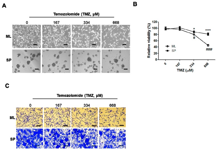
Figure 4.
Temozolomide (TMZ)-induced cell death in the ML and SP-forming conditions. Following TMZ treatment for 48 h in the ML or SP-forming conditions, cellular images were taken with an inverted microscope (A) and the chemosensitivity was measured using a viability assay kit as described in Materials and Methods (B); (C) Crystal violet staining was performed after treatment of increasing dose of TMZ for A172 cells in ML and SP-forming conditions. Spheres were re-attached on l-ornithine-coated plates. * p < 0.05 and *** p < 0.005 vs. 0 μM TMZ-treated ML, # p < 0.05, ### p < 0.05 vs. 0 μM TMZ-treated SP. Scale bars: 200 μm.

