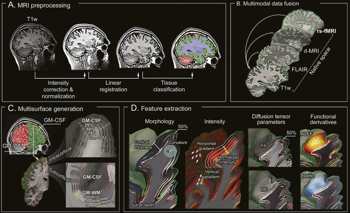
Figure 1. Image processing.
(A) T1-weighted MRI undergoes intensity nonuniformity correction and intensity normalization. T1-weighted images are linearly registered to the Montreal Neurological Institute 152 symmetric template and classified into tissue types. (B) Multimodal coregistrations map each modality to native T1-weighted space. (C) Models of the GM-WM and GM–CSF interface are generated, followed by reconstruction of equidistant intracortical and subcortical WM surfaces. (D) Features describing cortical and subcortical morphology, intensity, diffusion (FA, MD), and function are represented in a unified surface-based frame of reference. ALFF = amplitude of local functional fluctuation; d-MRI = diffusion-weighted MRI; FA = fractional anisotropy; FLAIR = fluid-attenuated inversion recovery; GM = gray matter; MD = mean diffusivity; ReHo = regional homogeneity; rs-fMRI = resting-state functional MRI.

