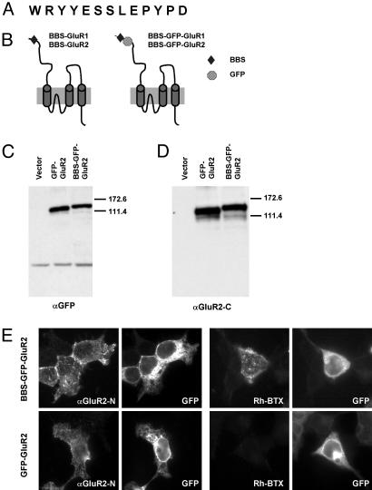
Fig. 1.
Expression of BBS-tagged receptor subunits in HEK 293 cells. (A) Amino acid sequence of the BBS. (B) Design of the BBS-tagged receptor subunits. BBS and GFP were inserted into the N-terminal extracellular region of the receptor subunits. (C and D) Western blotting of BBS-tagged subunits in HEK 293 cells. Both the GFP-GluR2 and BBS-GFP-GluR2 constructs were detected with anti-GFP (C) and anti-GluR2-C (D) antibodies. (E) Surface labeling of cells expressing BBS-GFP-GluR2 or GFP-GluR2 with anti-GluR2-N antibody (αGluR2N) or rhodamine-BTX (Rh-BTX). GFP, GFP signal detected in the cells. Both BBS-GFP-GluR2 and GFP-GluR2 constructs were expressed on the cell surface, whereas only the BBS-GFP-GluR2 construct was labeled with rhodamine-BTX on the cell surface.

