
Figure 5.
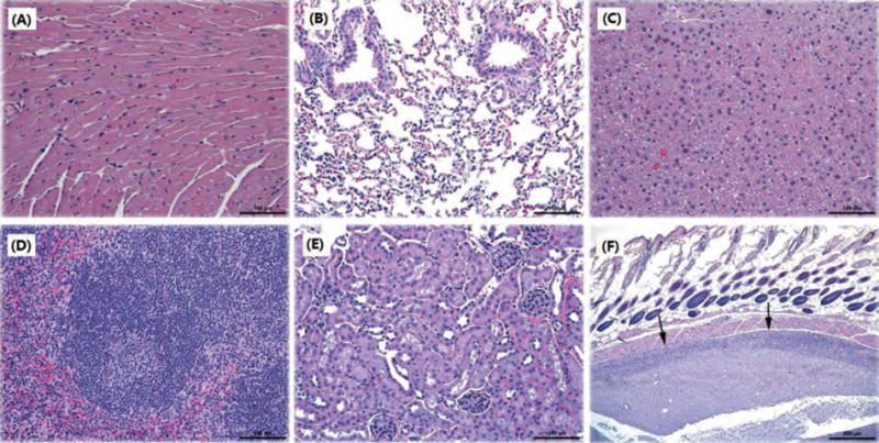
Representative histopathological images of different mouse tissues after subcutaneous administration of nicotine vaccines. No lesions were noted in (A) heart, (B) lung, (C) liver, (D) spleen, and (E) kidney. (F) is from a mouse given a vaccine with Alum. Arrows indicate subcutaneous lesions. Scale bars in (A)–(E) are 100 μm; for (F) the scale bar is 500 μm.