Figure 4.
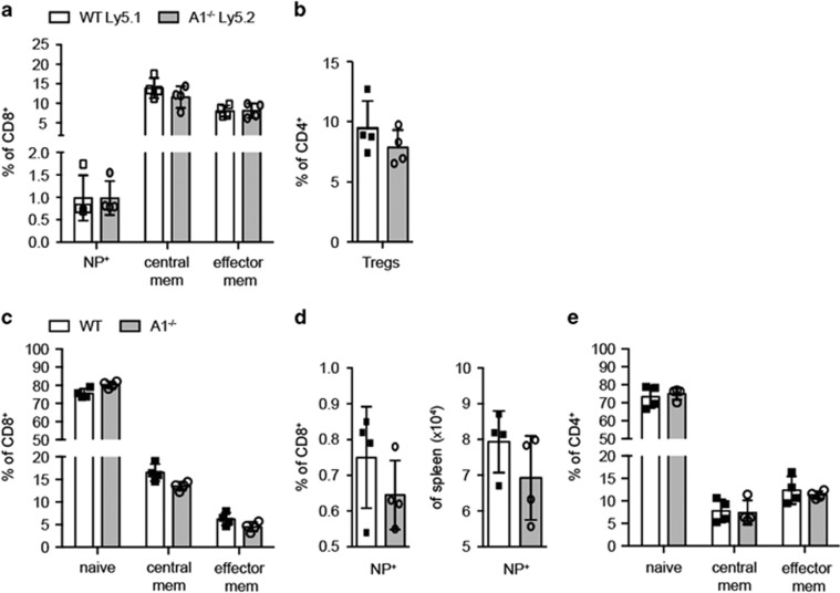
A1−/− T cells show normal in vivo memory formation. (a) 1 : 1 C57BL/6-Ly5.1 wild-type and C57BL/6-Ly5.2 A1−/− bone-marrow chimaeric mice were infected i.n. with 3000 pfu HKx31 influenza virus and spleen cells were analysed 41 days post infection by flow cytometry. CD8+ T cells were analysed for NP+ antigen-specific T cells as well as for central memory and effector memory-like phenotypes. (b) The frequencies of Treg cells within the CD4+ T cell population were analysed by intracellular flow cytometry for FoxP3 expression. (c) Wild-type and A1−/− mice were infected i.n. with 3000 pfu HKx31 influenza virus and spleen cells were analysed 41 days post infection by flow cytometry. CD8+ T cell populations were analysed for naive, central memory, and effector memory T cells. (d) The frequencies and total numbers of NP+ antigen-specific CD8+ T cells were assessed by flow cytometry. (e) CD4+ T cells in the spleens of infected mice were analysed for naive, central memory, and effector memory T cells. Bars represent means ±S.E.M. (n=4)

