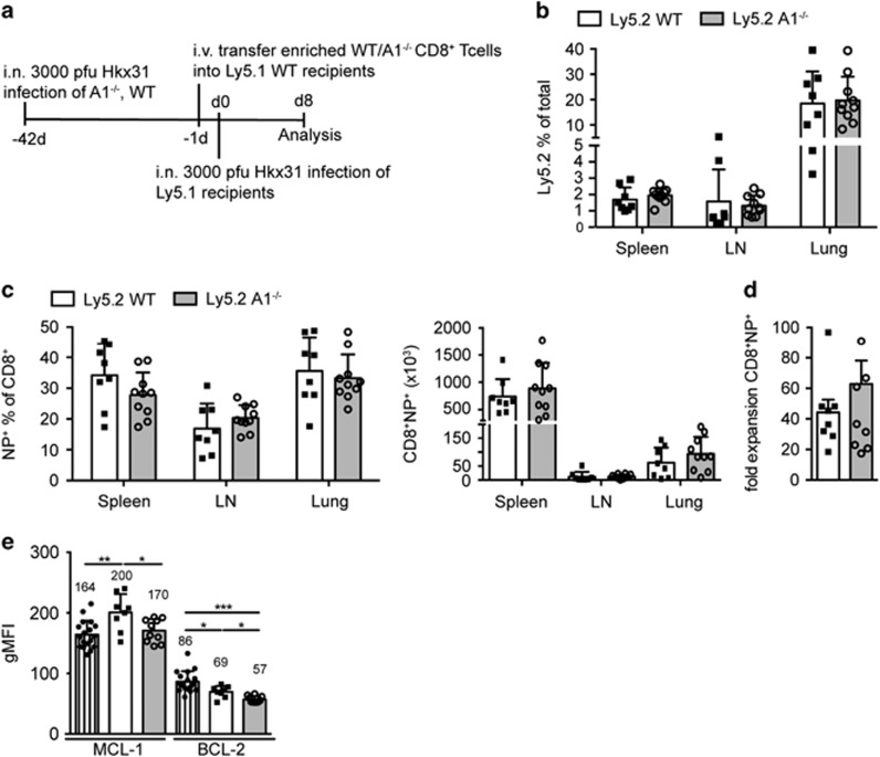
Figure 5.
A1−/− T cells expand normally in an influenza rechallenging model. (a) Schematic overview of the influenza recall immune response workflow: wild-type and A1−/− mice were infected i.n. with 3000 pfu HKx31 influenza virus and after 41 days mice were killed and CD8+ T cells were enriched by negative magnetic bead sorting. A total of 2 × 10⁴ NP-specific CD8+ T cells were transferred i.v. into naive C57BL/6-Ly5.1 wild-type recipient mice. One day later mice were infected i.n. with 3000 pfu HKx31 influenza and 8 days later lungs, mLN and spleens were isolated and analysed by flow cytometry. (b) The frequencies of Ly5.2+ donor cells were analysed in the spleen, mLN and lung infiltrates. (c) The frequencies and total numbers of NP+ antigen-specific Ly5.2+CD8+ T cells were analysed in the spleens, mLN, and lung infiltrates of infected mice. (d) The fold expansion of total Ly5.2+CD8+NP+ T cells was calculated by comparing the numbers of injected Ly5.2+CD8+NP+ T cells to the number of recovered Ly5.2+CD8+NP+ T cells from spleen, mLN and lung. (e) Intracellular staining for MCL-1 and BCL-2 was performed on total spleen cells of infected mice and the geometric mean fluorescent intensity (gMFI) for MCL-1 and BCL-2 protein in Ly5.2+CD8+ donor T cells was compared with that from similarly stained Ly5.1+CD8+ host T cells. Bars represent means±S.E.M. (n=8–10). A Mann Whitney statistical test was performed, *P<0.05, **P<0.01, ***P<0.0001

