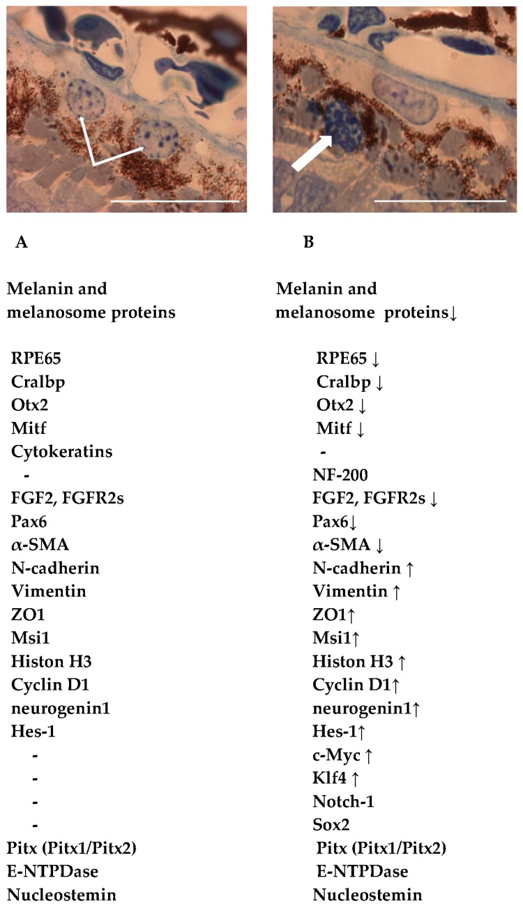
Figure 1.
Accumulated data on morphological and molecular features of native retinal pigment epithelium (RPE) cells and those at the beginning of natural reprogramming to neuronal and glial cells of regenerating retina [6,7,11,12,13,14,17,18,19,20,21,22,23,24]. (A) RPE cells (thin white arrows) in the RPE layer of the newt Pleurodeles waltl; (B) RPE cell that left its layer and stays at the beginning of reprogramming (thick white arrow); Scale bar: 100 µm. See details in the text. Down- (↓) and up- (↑) regulation of gene/protein expression.

