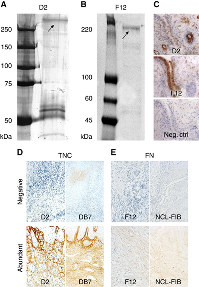
Figure 1.
Characterisation of the new matrix antibodies. Coomassie-stained SDS-7.5% PAGE of the target antigens from the cultured hMSC lysate immunoprecipitated using the new anti-tenascin-C monoclonal antibody D2 (A) or anti-fibronectin monoclonal antibody F12 (B). Molecular weight markers are shown on the left of the gels: Bio-Rad Dual Colour (A) and Sigma Colorburst (B). The identified target antigen is marked with arrows. Bands migrating at ∼60 kDa region are mouse antibody fragments detached from the resin (A). The immunostaining of the synovia tissue sections with anti-tenascin-C D2, anti-fibronectin F12 and idiotype IgG1 as negative control (C). New antibodies were compared with commercial ones by staining representative samples from ‘negative' and ‘abundant' groups using both D2 and commercial DB7 for TNC (D), and both F12 and commercial NCL-FIB for FN (E).

