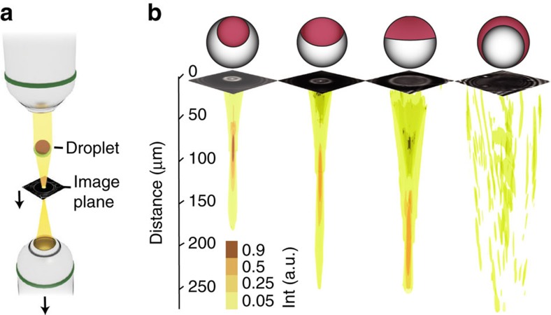
Figure 2. Variable focusing.
(a) Schematic of the setup used to record the light field behind the droplets. The bottom objective scans through the z-direction. (b) Iso-surfaces of the reconstructed light fields behind the droplets for different internal droplet morphologies. Representative image data sets captured immediately behind the droplets show slices of the scan, from which the light field was reconstructed. Droplet sketches are offset upwards by 10 μm to not obstruct slice images.

