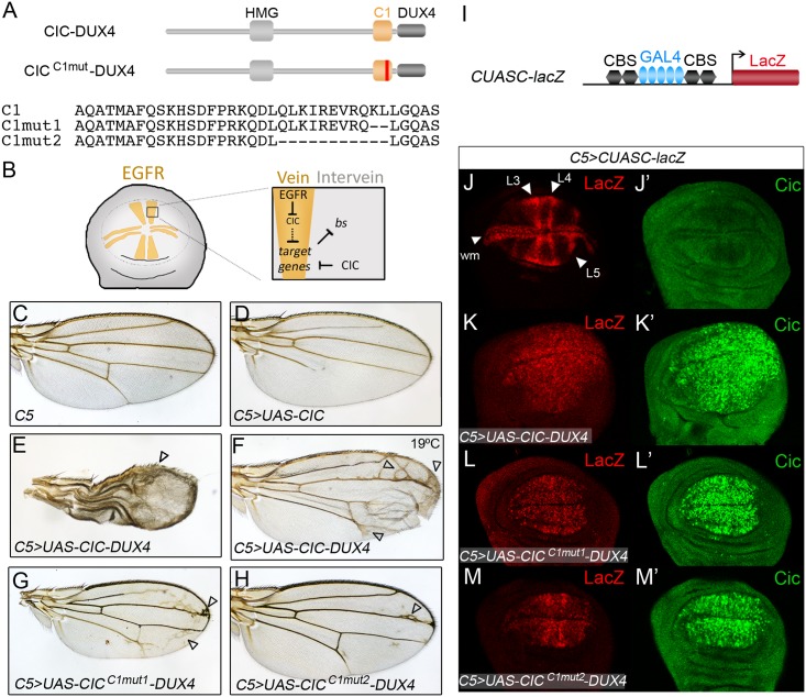
Fig 7. The C1 domain is required for the activity of a CIC-DUX4 fusion in the Drosophila wing.
(A) Diagram of CIC-DUX4 chimeras expressed in the wing using the GAL4-UAS system. CICC1mut-DUX4 indicates two different derivatives carrying CRISPR-Cas9-induced mutations (vertical red line) in the C1 domain; partial sequences of intact and mutant C1 domains are shown below. (B) Model of CIC function in the wing primordium. The pattern of wing veins is established in the imaginal disc through localized activation of the EGFR signaling pathway, which downregulates CIC in presumptive vein cells (yellow). CIC in turn promotes the intervein fate by repressing (directly or indirectly) EGFR-induced genes such as ventral veinless and decapentaplegic, while indirectly maintaining blistered (bs) expression in intervein cells [7,12,67]. (C-H) Wing phenotypes induced by expression of CIC (D), CIC-DUX4 (E, F) and two CIC-DUX4 mutant derivatives carrying deletions in the C1 domain (G, H) under the control of the C5 GAL4 driver; a control wing with GAL4 driver only is shown in C. Arrowheads indicate broadened veins and ectopic vein material in CIC-DUX4-expressing wings. Unless otherwise indicated, all panels were obtained by raising flies at 25°C; panel F shows the weaker phenotype resulting from induction of CIC-DUX4 at 19°C. (I) Diagram of the CUASC-lacZ reporter driven by a synthetic enhancer composed of five GAL4 binding sites flanked by two CBSs on either side. (J-M’) Late third-instar wing discs doubly stained with anti-lacZ (J-M) and anti-Cic antibodies (J’-M’). J and J’ show a wild-type disc carrying the CUASC-lacZ reporter. K-M’ show representative discs expressing intact and mutant CIC-DUX4 proteins using the C5 driver, which is expressed in the wing pouch and serves to activate both the effector genes and the CUASC-lacZ reporter.

