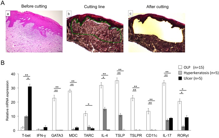
Fig 5. mRNA expression of Th-related cytokines, chemokines, and transcription factors in the selective BM specimens by using Laser Microdissection (LMD).
A, Hematoxylin and eosin (HE)-stained BM specimens (a). The infiltration of inflammatory cells was indicated by a green line (b). The preselected area was extracted by using a guided laser beam after LMD. (c). Scale bars, 500 μm. B, mRNA expression levels of Th-related cytokines, chemokines, and transcription factors in BM samples from patients with OLP (n = 15), hyperkeratosis (n = 5) and ulcer (n = 5). mRNA expression levels of Th-related cytokines, chemokines, and transcription factors were estimated quantitatively as described in the Methods section. Statistical significance of differences between groups was determined by Kruskal-Wallis tests (**P < 0.01, *P < 0.05).

