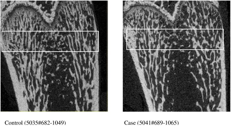
Fig 2. Coronal section micro-CT images of ultra-distal femurs of left (non-paretic) legs of animals with sham surgery control and middle cerebral occlusion stroke (case).
White boxes indicate the regions of analysis for bone density and bone micro-structural parameters. Image on left = Control (5035#682–1049). Image on right = Case (5041#689–1065).

