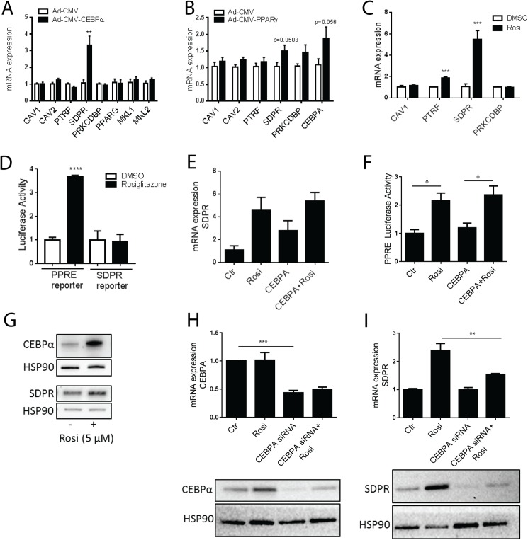
Fig 3. Overexpression of CEBPα and PPARγ activation increase SDPR expression in primary adipocytes.
Panels A and B show qPCR analysis of caveolin and cavin expression in primary rat adipocytes following 20–24 h of incubation with adenovirus encoding CEBPα (Ad-CMV-CEBPα) (A), PPARγ (Ad-CMV-PPARγ) (B) or control adenovirus (Ad-CMV-null). n = 6 independent experiments. Adipocytes were incubated with Rosiglitazone (5 μM) or vehicle (DMSO) for 20–24 h, and the relative expression of CAV1, CAV2, PTRF, SDPR, and PRKCDBP analyzed using qPCR (C, n = 4 independent experiments). In panel D (left bars), adipocytes were transfected with PPRE luciferase reporter and Renilla reporter plasmids, followed by overnight (20 h) incubation with Rosiglitazone (5 μM) or vehicle (DMSO). Luciferase activity was measured in cell lysates using a luminometer, and the values normalized to Renilla luciferase activity. Each sample was measured in triplicate (n = 3 independent experiments). Adipocytes were also transfected with an SDPR promotor reporter (right bars) and incubated overnight (20 h) with Rosiglitazone (5 μM) or vehicle (DMSO). Cell lysates were subjected to luciferase activity assay and each sample was measured in triplicate. n = 3 independent experiments. Adipocytes were incubated with DMSO (ctr), CEBPα adenovirus, Rosiglitazone (5 μM), or their combination overnight (20 h), and analyzed using qPCR (E, n = 3 independent experiments). In panel F, adipocytes were transfected with PPRE luciferase reporter and Renilla reporter plasmids, followed by overnight (20 h) incubation with adenovirus encoding CEBPα, Rosiglitazone (5 μM) or vehicle (DMSO) as indicated. Cell lysates were subjected to luciferase activity assay and each sample was measured in duplicate. n = 3 independent experiments. Western blot showing CEBPα and SDPR in cell lysates from primary adipocytes incubated with or without Rosiglitazone 20 h (DMSO, ctr). HSP90 was used as loading control. (G). In H, CEBPA was silenced using siRNA (or scrambled (ctr)) in 3T3-L1 cells on day 4 of differentiation. At day 6, cells were stimulated with Rosiglitazone (1 μM) for 20 h. n = 3 independent experiments, each sample run in duplicate. CEBPα silencing was confirmed by western blotting, representative image is shown below graph (H). mRNA expression of SDPR following CEBPA silencing is shown in I. Protein level of SDPR shown below graph (I). HSP90 used as loading control (H and I). Data is presented as means±SEM, *p≤0.05, **p≤0.01, ***p≤0.001, and ****p≤0.0001.

