
Figure 3.
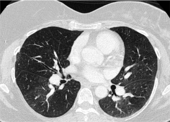
Chest CT angiogram demonstrating marked mosaic attenuation in bilateral lung fields as well as pruning of distal pulmonary arteries and enlargement of proximal pulmonary arteries.
Official websites use .gov
A
.gov website belongs to an official
government organization in the United States.
Secure .gov websites use HTTPS
A lock (
) or https:// means you've safely
connected to the .gov website. Share sensitive
information only on official, secure websites.
Chest CT angiogram demonstrating marked mosaic attenuation in bilateral lung fields as well as pruning of distal pulmonary arteries and enlargement of proximal pulmonary arteries.