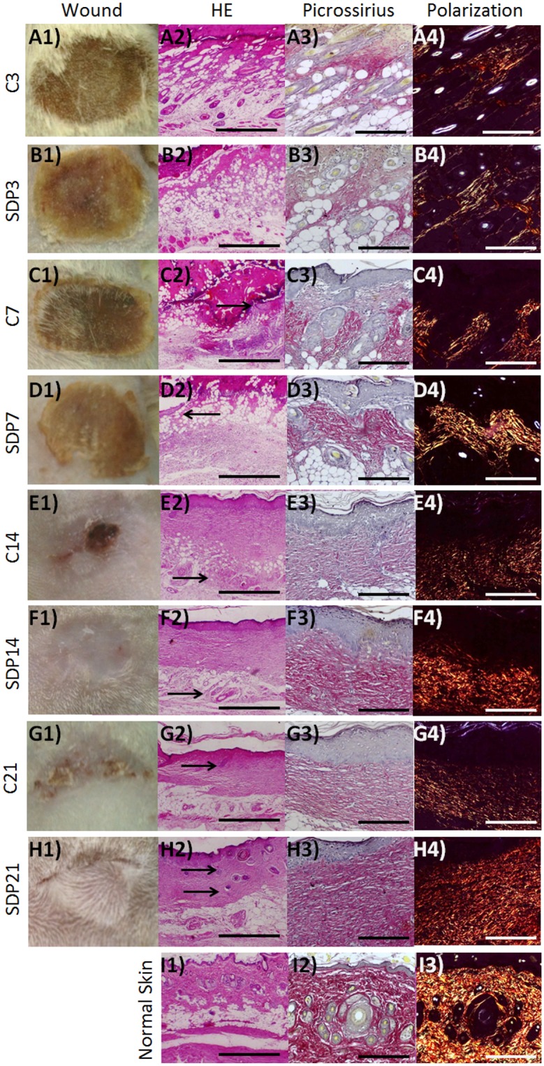
Fig 1. Macroscopic and histopathologic analysis of the healing process in a second degree burn.

(a) C3 and (b) SDP3, inflammation phases, with leukocytes influx and presence of adipocytes in both groups. (c) C7 and (d) SDP7, proliferation phase with re-epithelialization (arrows). Up to 7 days weakly stained collagen fibers (a3, b3, c3 and d3) and low birefringent after polarization (a4, b4, c4 and d4) in the dermis of both groups. (e) C14 and (f) SDP14, complete reepithelialization and fast healing in SDP 1% group (e1 and f1). Dermis and hypodermis (e2, f2—arrows) are better organized and collagen fibers more strongly stained in the group treated with SDP 1% (e3, f3). (g) C21 and (h) SDP21, remodeling phase, the epithelial attachments (g1, h1, g2, h2—arrows) may be observed in larger amount and with increased birefringence under polarized light in the groups treated with SDP 1% (g4 and h4). Normal Skin (i). (a1, b1, c1, d1, e1, f1, g1, h1 and i1) Macroscopic analisys. (a2, b2, c2, d2, e2, f2, g2, h2 and i2) Staining: HE. Bars 500 μm. Picrossirius staining without (a3, b3, c3, d3, e3, f3, g3, h3 and i3) and after (a4, b4, c4, d4, e4, f4, g4, h4 and i4) polarization. Bars 200μm.
