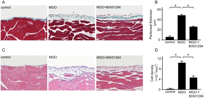
Fig 2. BIX01294 suppresses peritoneal thickening and cell density in MGO-injected mice.
(A) Masson’s trichrome staining shows the typical thickness of peritoneal tissue in control mice, MGO-injected mice, and MGO-injected mice treated with BIX01294 (original magnification, ×200). (B) Graph indicates quantification of the peritoneal thickness in the three groups of mice. (C) Hematoxylin-eosin staining shows typical cellularity of peritoneal tissue in control mice, MGO-injected mice, and MGO-injected mice treated with BIX01294 (original magnification, ×200). (D) Graph indicates quantification of cell density in the three groups of mice. Data are expressed as the mean ± SE. Statistical analysis was performed by analysis of variance followed by Tukey’s post-hoc test. *P < 0.05, n = 5 mice per group.

