
Fig 3. Viral loads in the tissues following necropsy of ZIKV-infected rhesus macaques.
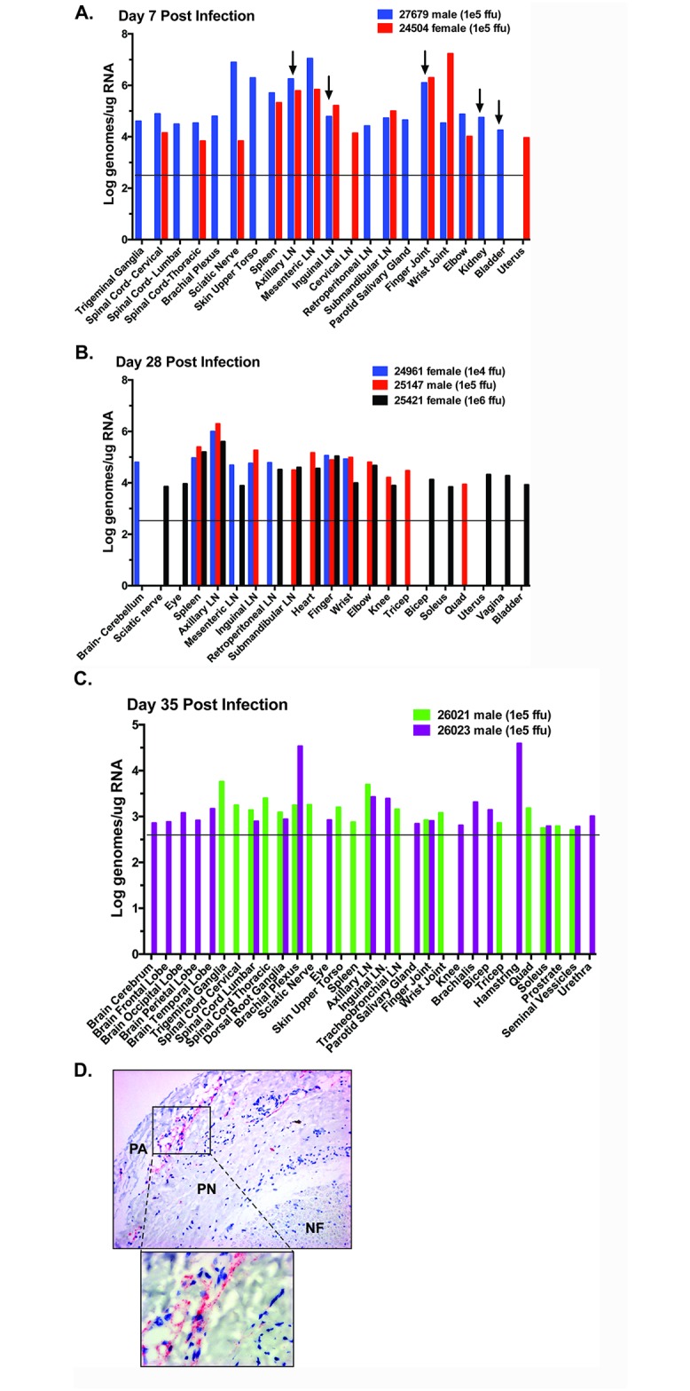
One-step qRT-PCR was used to measure ZIKV RNA loads in the tissues of animals in Cohort 2 (A), Cohort 1 (B), and Cohort 3 (C). Total RNA was generated using the Trizol method on precleared samples following bead beating. Approximately 80 different tissues were assessed for the presence of viral RNA. Shown are the tissues with positive detection in at least one of the animals per cohort. Arrows indicate samples in which virus was successfully co-cultured from tissue homogenate. Approximate limit of detection at 1e4 genomes/ml is based on a detection limit of ~100 genomes in each reaction (S1 Fig) as indicated by the horizontal line. (D) Paraffin sections of sciatic nerve cut in cross section were hybridized with ZIKV specific chromogenic probe (red) and counterstained with hematoxylin (blue). Nerve fibers (NF) show a normal distribution within the endoneurium surrounded by perineurium (PN) and perineurial adventia (PA). Hybridization for ZIKV was robust but limited to the PA region. Original magnification was 50X.