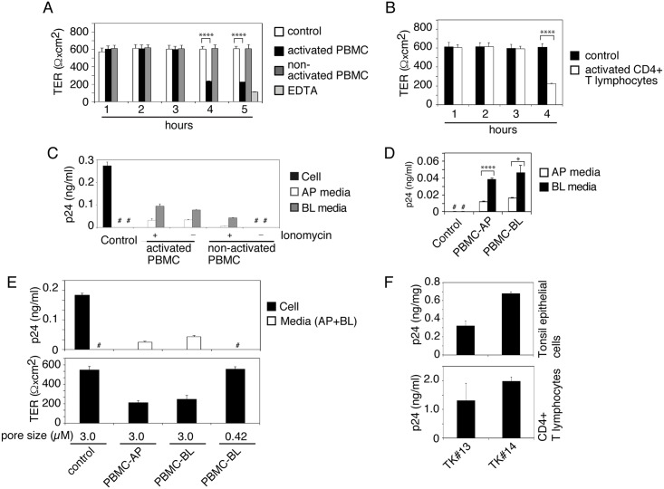
Fig 5. Cocultivation of activated PBMC and CD4+ T lymphocytes with epithelial cells containing HIV-1 induces virus release.
(A) Activated and nonactivated PBMC were added to the AP surface of polarized tonsil epithelial cells (2:1), and after 1, 2, 3, 4 and 5 h the TER was examined. As a control, one set of polarized tonsil cells were not cocultivated. Another set of cells were treated with 10 mM EDTA for 30 min. (B) Activated CD4+ T lymphocytes were added to the AP surface of tonsil epithelial cells, and after 4 h the TER was examined. (A, B) Mean values ± SEM of three independent experiments (in triplicate) are shown (n = 3). ****P < 0.00001, TER of polarized cells cocultivated with activated PBMC or CD4+ T lymphocytes compared with TER of control cells. (C) Activated and nonactivated PBMC with or without ionomycin were added to the BL surface of tonsil cells containing intracellular HIV-1SF33 at day 6. After 4 h of cocultivation AP and BL medium was collected separately, centrifuged, and supernatants were examined for released virus using ELISA p24. Polarized epithelial cells and culture medium without cocultivation with PBMC were used as a control. (D) Activated PBMC were added to the AP or BL surface of tonsil cells containing HIV-1. Four hours later, AP and BL medium was examined for HIV-1 p24. (E) Polarized tonsil epithelial cells were grown in Transwell inserts with 3-μm or 0.42-μm pore size and exposed to HIV-1SF33 from AP membranes. Epithelial cells with sequestered virus at day 6 were cocultured with activated PBMC from the AP surface of polarized cells grown in inserts with 3.0-μm pore size and from BL membranes in inserts with 3-μm or 0.42-μm pore size. After 4 h, the TER of polarized cells was measured, and the culture medium from AP and BL membranes were combined for ELISA p24. (F). Matching tonsil keratinocytes and CD4+ T lymphocytes were isolated from the same tonsil of 2 independent donors. HIV-1SF33 was added to the AP surface of polarized tonsil epithelial cells, and at day 6 intracellular virus was examined by ELISA p24 (F, upper panel). One set of cells containing virus were used for cocultivation of autologous CD4+T lymphocytes. After 4 h, lymphocytes were collected, grown for 12 days and examined by ELISA p24 (F, lower panel). Data in panels C-F represent one of two or three independent experiments and are shown as mean ± SEM of triplicate values. (D) *P < 0.01, ****P < 0.00007, BL release of virus compared with AP release.

