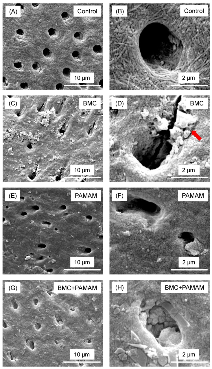
Figure 5.
Representative SEM images of demineralized root dentin surface perpendicular to tubule axis after 21 days cyclic artificial saliva/lactic acid regimen: (A,B) control group, (C,D) BMC group, (E,F) PAMAM group, and (G,H) BMC + PAMAM group. Left column is at a lower magnification. Right column is at a higher magnification. Exposed collagen fibrils were detected in (A,B). (C–F) showed that regenerated minerals precipitated on the root dentin surface. Arrows showed minerals partially occluding the dentinal tubules. In (G,H), the dentin was covered by the remineralized mineral crystals.

