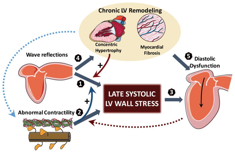
Figure 1. Working pathophysiological model linking time-varying MWS and diastolic dysfunction.
Arterial wave reflections selectively increase mid-to-late systolic LV load and MWS (1). However, the effect of wave reflections on MWS is strongly influenced by the LV contraction pattern. Normally, brisk force development and fiber shortening occur in early systole, resulting in LV ejection and a dynamic reconfiguration of LV geometry that results in a mid-systolic reduction in MWS relative to LV pressure, thus protecting the cardiomyocytes against excessive load in mid-to-late systole (a period of increased vulnerability). The phenomenon of shortening-deactivation, which decreases force development after rapid shortening, and increases the relaxation rate of muscle, is a perfect “match” to this normal physiology, because sustained myocardial force development in mid-to-late systole is unnecessary to preserve fiber shortening and LV ejection against the load imposed by wave reflections. However, in the presence of contractile abnormalities that compromise early ejection (and reduce EF1) (2), the mid-systolic dynamic geometric reconfiguration of the LV that favors a reduced MWS relative to pressure does not occur. Late systolic MWS therefore more directly correlates with LV pressure, indicating that the myocardium is less protected from late systolic arterial load induced by wave reflections, thus facilitating their ill effects (curved blue solid arrow). In these conditions, shortening-deactivation does not operate fully, such that force development continues or increases in mid-to-late systole, tending to preserve the overall EF on one hand, but impairing relaxation (3) on the other. Late systolic LV load from wave reflections also promotes chronic LV hypertrophy/fibrosis11,12 (4), which can induce diastolic dysfunction (5) and contractile abnormalities (curved blue dotted arrow) in their “own right”. Concentric LV hypertrophy/remodeling is also intrinsically associated with a blunted mid-systolic shift in the pressure-MWS relation, facilitating a myocardial loading sequence characterized by increased late systolic load (curved red solid arrow). Finally, reduced lengthening-activation from increased ventricular stiffness may impair early systolic contraction (curved red dotted arrow). For simplicity, the potential role of other factors (stored potential energy in the myocardium/restoring forces, interventricular dependence, pericardial restraint, ascending aortic stretch and

