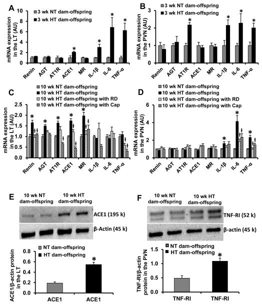
Figure 2.
Quantitative comparison of the mRNA expression of renin-angiotensin system components and proinflammatory cytokines in the lamina terminalis (LT) and paraventricular nucleus (PVN) in male offspring of normotensive (NT) and hypertensive (HT) dams at 3 weeks of age (Fig 2A and 2B) and at 10 weeks of age before and after renal denervation (RD) and captopril (Cap) treatment (Fig. 2C and 2D). Fig. 2E and 2F show representative Western blots and quantitative comparison of protein levels for ACE1 in the LT (Fig 2E) and TNF-α receptor I (TNF-RI) in the PVN (Fig 2F) in male offspring of NT and HT dams at 10 weeks of age. Values are corrected by β-actin and expressed as mean ± SEM. (n=5/group; * p<0.05 vs NT-dam offspring; ƚ p<0.05 vs. HT-dam offspring without RD or Cap treatment).

